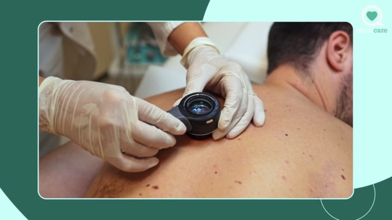
How is skin cancer diagnosed?
Watch as dermatologist Dr. Ellen Marmur discusses how skin cancer is diagnosed.
Transcript
So this is the most important part of the diagnosis step is actually taking a skin biopsy. It sounds horrible.
It's not that bad. [MUSIC PLAYING]
Skin cancer is diagnosed in several ways. First of all, we need to detect it. So really first of all, you can help diagnose it
by detecting anything new on your skin or anything that's changing. That helps a lot. If I have a patient come in and say to me this spot bothers me
and I look at it and I say it looks OK to me-- if they are bothered by it, I'm going to look twice and even
three times very closely to check it out. We have these new gadgets now in the dermatologist's office. And not only do we have a big magnifying glass or something
called a Wood's lamp, which is the old disco black light thing. It helps show pigment better. We now have a gadget called a dermatoscope.
And a dermatoscope is using a polarized, small handheld device to look up close at the different types of pigment
of a mole. And that helps us a lot whether or not if we're going to biopsy something.
So this is the most important part of the diagnosis step is actually taking a skin biopsy. It sounds horrible.
It's not that bad. They give you a tiny, tiny shot of lidocaine, which is like novocaine. It numbs the skin instantly.
And that feels like a little mosquito bite. So that's about all you should feel. We wait about 10 seconds. And then we take a small sample of whatever
spot looks suspicious. You shouldn't feel anything. We stop the bleeding and put on a Band-Aid. And that should heal up between within one to two weeks.
So it's not a big deal to have a biopsy. We send that to a histopathology laboratory
where they look under the microscope and look at the architecture of the cells and tell you whether or not those are abnormal or normal cells.
That process can take two days, up to two weeks if they need to do special stains. So you should have your diagnosis
within one to two weeks of having had your biopsy. If it is a skin cancer, there are different ways to remove it.
Usually, if it's on an important area of the body and if it's a basal cell cancer or a squamous cell cancer,
we do a technique that I specialize in called Mohs surgery. M-o-h-s is the name of the guy who invented the surgery, OK.
And this is a way of doing almost a fancy biopsy while you wait. We remove the spot. We make a map out of it, bring it into the laboratory,
and we check 100% of those margins. Imagine peeling an orange. We're looking at 100% of the margin of that tissue
to be sure that the skin cancer doesn't have any roots that are left behind. If there are roots, we take a second little stage
of surgery, same process, took it under the microscope. So you're waiting around for a couple of hours for the day. But you know that your skin cancer is out.
And then we sew it up in what's called reconstructive surgery. This is all done under local lidocaine. It's extremely safe.
If you have a dysplastic nevus, which is an atypical brown spot or a melanoma,
we don't use the rapid staining in my Mohs lab. We do a standard surgery where we take four-millimeter margins
up to a centimeter margins, depending on what the lesion is. And we remove that spot and put it back together right away
with stitches. [AUDIO LOGO]
skin cancer
Browse videos by topic categories
A
B
C
D
E
F
G
H
I
J
K
L
M
N
O
P
Q
R
S
T
U
V
W
X
Y
Z
ALL