Updated on October 11, 2023.
Of all your senses—sight, smell, touch, taste, and hearing—eyesight is arguably your most important. It not only helps you appreciate a beautiful sunset but also navigate the world each moment of the day. Clearly, something so important deserves special attention and good care. And that's where a healthcare provider (HCP) with expertise in eye care comes in.
So who should you see? And when should you go? And just as important, what can you expect when you get there?
What kind of eye provider should I see?
For many of us, the "who" part is simple. We go to whatever eye care provider is covered by our insurance plans. But even within a healthcare plan, you often have choice.
How often should I go?
How often you see an eye care professional depends on your age, medical conditions, and other factors, such as your eye health history. So work with your personal eye care specialist for a schedule that fits your needs. As a baseline, all people should have an eye disease screening by age 40.
What happens at my eye exam?
So what's likely to happen when you finally get your time with the HCP? Here are six key components of a vision exam:
Medical history
A screening should start with an overview of your medical history. Your eye care professional will want to know about any medical conditions you have and about any medications or supplements you're currently taking. You should also describe any vision problems that you have currently, as well as any history of eye problems in your blood relatives.
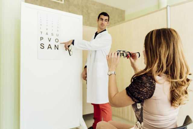
Visual acuity check
Next, the HCP will likely check your visual acuity—how clearly you're able to see. You'll be asked to read characters off an eye chart that's 20 feet away while wearing your prescription glasses or contacts. The HCP will check the vision in each eye while the other one is covered.
The test results are reported as a fraction. Someone with 20/20 vision can see characters from 20 feet away, which is considered normal. A bigger bottom number means worse vision. Someone with 20/100 vision can see characters from 20 feet away as clearly as someone with normal vision can see from 100 feet away.
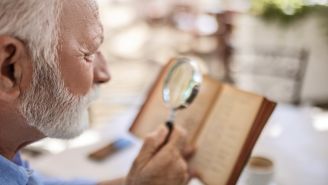
Your HCP may also check how well you read things up close, especially if you're over 40, because the risk of presbyopia (difficulty seeing up close) increases as we age.
Refraction test
If your vision seems off, your eye care professional will give you a refraction test to see if glasses or contacts (or changes to your existing prescription) could help you see more clearly. This involves reading an eye chart while looking through different lenses and letting your HCP know whether the view gets fuzzier or clearer when they change the lenses. They'll continue to adjust the lenses until refractive errors are corrected and your vision is sharp.
Checking for disease
Your HCP may also examine your peripheral vision and central vision, checking for signs of glaucoma or problems with your maculas (the part of your eye that produces central vision).
To test your peripheral vision, your HCP might ask you to cover one eye and stare at a fixed point while they move their hand a few feet away from your face until you see it. Or they may use a more sophisticated test that involves looking into a special device and pressing a button every time you see a flashing light. For central vision testing, your HCP will probably ask you to look at an Amsler grid (a square consisting of a grid pattern with a dot in the middle) and ask if you see any blurry, wavy, or missing lines.
Checking eye muscles and function
Your eyes have a lot of moving parts that join together to help you see, so your HCP will want to know how well all the bits and pieces of your eyes are working. To do this, they may ask you to follow an object (usually a pen or a light) with your eyes as you keep your head still. They'll note how your eyes move up, down, left, right, and toward the tip of your nose. Good results mean your extraocular muscles are working well. Your eye care professional might also cover and uncover each eye as you look at an object to see how well your eyes work together.
Dilation and inner and outer structures
And no eye exam would be complete without taking a peep at your pupils. In a dimly lit room, the HCP will see how your pupils dilate as they shine a beam into each eye. Healthy pupils dilate and constrict in response to how much light they receive. A handheld instrument called an ophthalmoscope will allow the HCP to take a closer look at the inner structures of your eyes, such as the retina, choroid, optic nerve area, and macula. Sometimes, eye drops that affect your vision for a while are used with this scope as well.
Finally, the outer parts of your eyes will be examined for any signs of injury, infection, or inflammation.
Special Tests
Depending on your medical history and exam results, you might need additional tests that aren't included in a standard eye exam. Here are a few examples:
Slit-Lamp Exam
This microscope allows for a better look at the structures in the front of your eye: the sclera, conjunctiva, cornea, iris, and lens. Your HCP may also use an orange dye called fluorescein to help detect injuries on the colorless cornea.
Color Vision Test
Images composed of colored dots can help reveal if you have trouble distinguishing between certain colors.
Tonometry
This test checks your intraocular pressure, important for diagnosing glaucoma. A puff of air is delivered to the eye to flatten your cornea, and the energy delivered is linked with the pressure in your eyes. Or your HCP might give you numbing eye drops and use instruments that lightly touch your corneas to measure pressure.
Before you leave
Did you get all the tests you were expecting? What were the results? Did you have questions about any of them? Before you leave your appointment, make sure you understand where things stand with your eyes.